What the component does
A pharmaceutical particle graticule offers method-specific comparison features in the eyepiece. It is a reference component, not a complete particle-testing system.

Support calibrated visual particle comparison within a defined pharmaceutical microscopy method.
The calibration and inspection must use the optical setup required by the chosen method.
The outlines illustrate comparison only; their dimensions and interpretation must follow the applicable method.
A pharmaceutical particle graticule offers method-specific comparison features in the eyepiece. It is a reference component, not a complete particle-testing system.
Reference artwork, sample preparation, illumination and magnification need to match the selected procedure. Verify the stage-to-eyepiece conversion before use.
A similar-looking pattern is not necessarily interchangeable. The presence of a graticule alone does not establish compliance of the instrument or the test result.
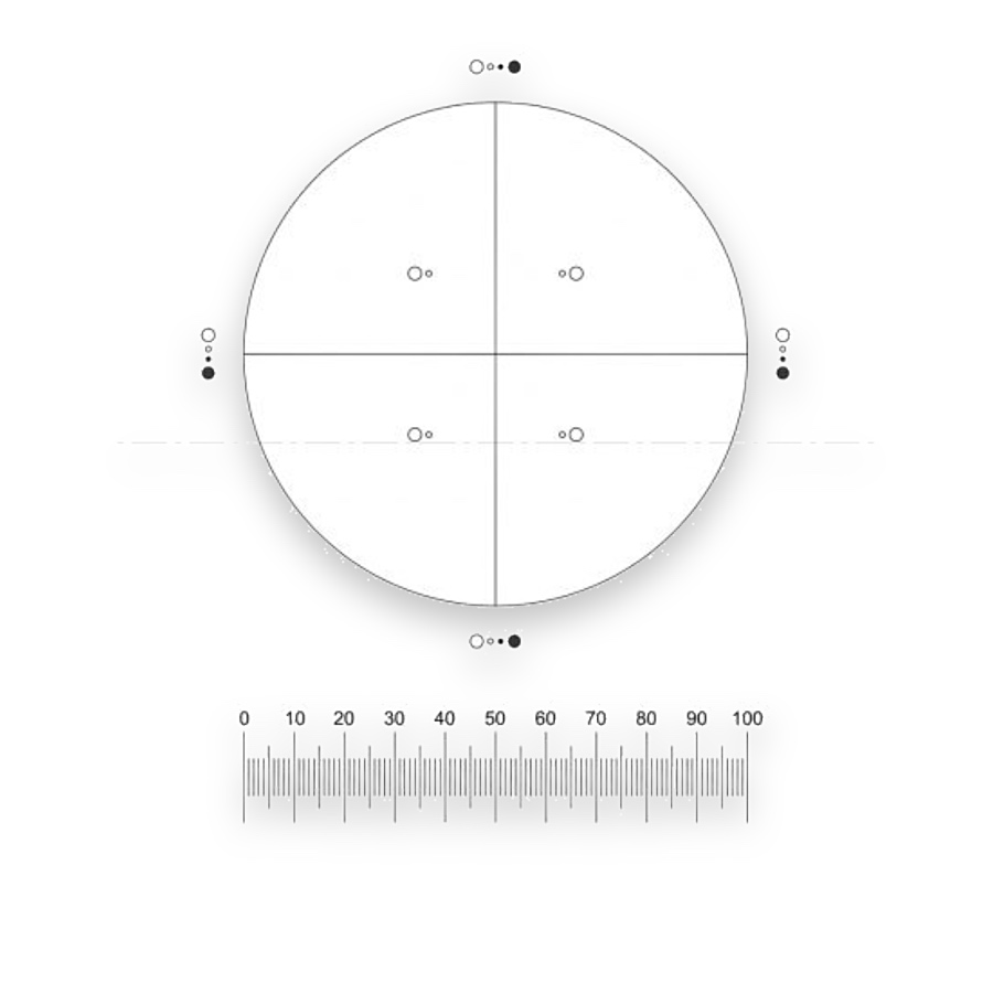
Counting-field and size-comparison pattern for the specified microscopy method.
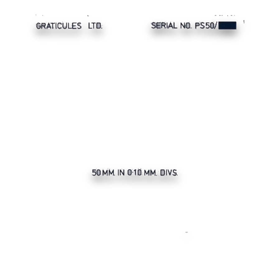
Length reference for microscope calibration.
Microscopic particle examination uses a prepared sample, defined observation conditions and a counting rule to classify visible particles. A suitable eyepiece reticle provides the field and size-comparison features needed by that procedure. It forms one part of the measurement apparatus.
Select the pattern specified by the applicable method, then calibrate its projected size in the microscope. Reference to USP <788> describes a method context, not a blanket claim that any similarly named reticle or complete instrument meets that method.
A general counting grid subdivides area but may not supply the comparison features required at a particular particle threshold. The intended reticle must give the operator the correct visual reference at the working magnification.
Sample preparation, illumination, focus, magnification and counting conventions all influence results. Use the scale reference to verify the viewing configuration and retain the applicable procedure with the observation records.
Pharmaceutical particle microscopy uses method-specific fields and size-comparison references.
Match the pattern to the adopted procedure and working magnification, then verify its projected scale with a length reference.
A grid divides the field but may lack the required size-comparison features. Choose the actual artwork and optical configuration for the method.