What the component does
The eyepiece reticle provides a visible scale. The stage micrometer supplies the known physical length used to calibrate that scale; they occupy different optical planes.

Turn microscope divisions into a reliable physical length by calibrating the observation system before measuring a specimen.
Align the imaged stage scale with the eyepiece divisions and calculate length per division for this optical configuration.
Multiply the observed division count by the calibrated length per division. Recalibrate after changing the objective or optical magnification.
The eyepiece reticle provides a visible scale. The stage micrometer supplies the known physical length used to calibrate that scale; they occupy different optical planes.
Record the objective, eyepiece and any intermediate magnification with the conversion factor. An XY micrometer can check both image directions when needed.
Using a conversion factor from another objective creates a systematic size error. Thick or poorly focused reticle lines also make endpoints harder to judge.
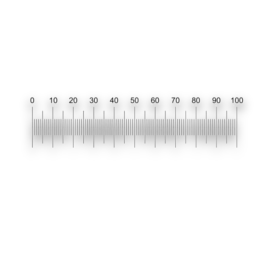
Graduations for calibrated visual length measurement.
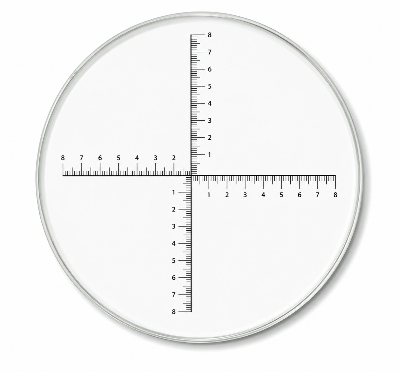
Orthogonal graduations for comparing X and Y dimensions in the same field.
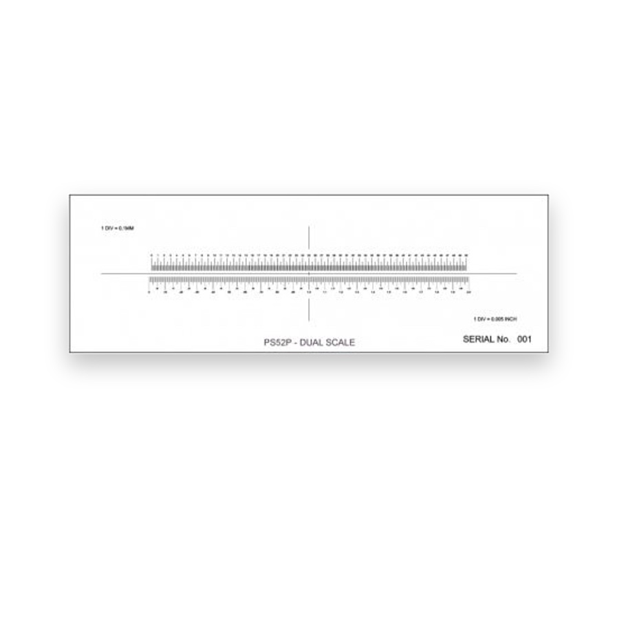
A length reference for pixels or eyepiece divisions.
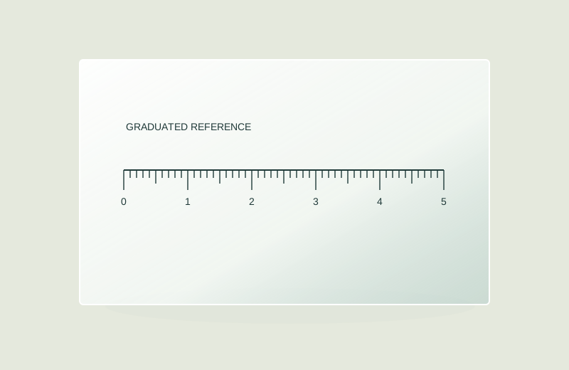
Select one-axis or XY specimen-plane calibration scales.
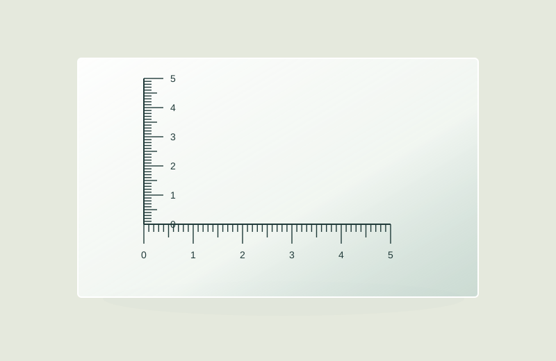
Two-axis scale and alignment checks.
Select measurement and counting patterns for an eyepiece.
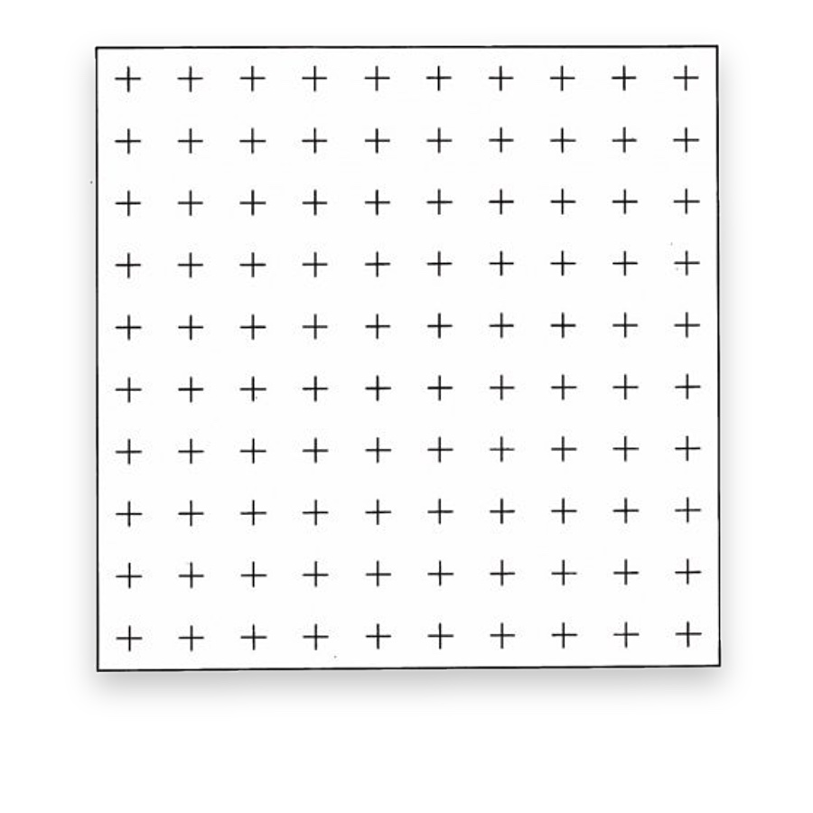
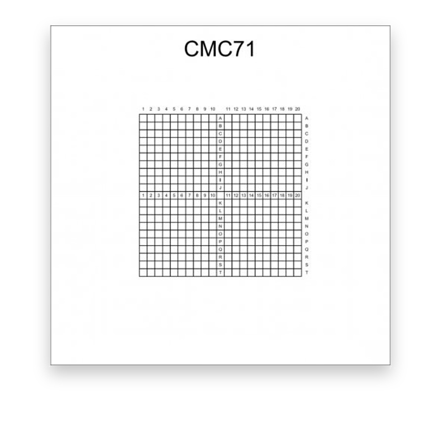
Field subdivisions for systematic counting.
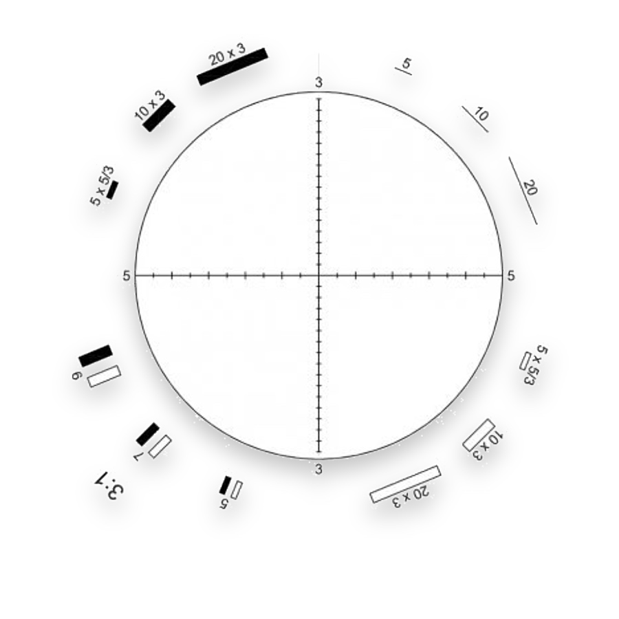
Circular reference fields for compatible fibre-counting methods.
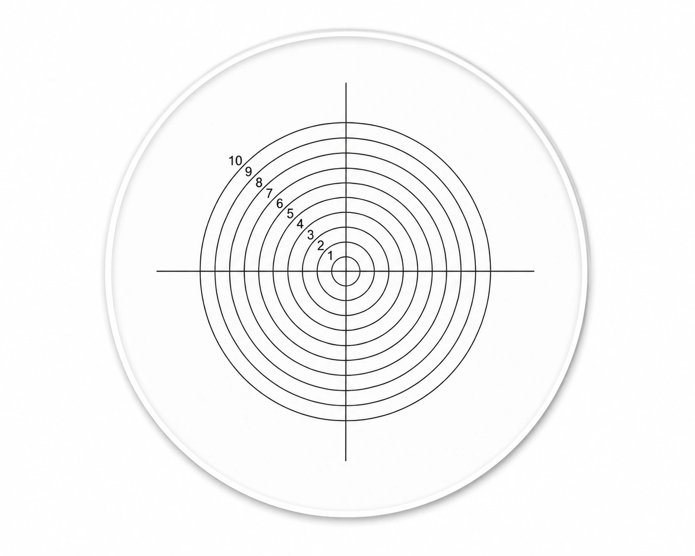
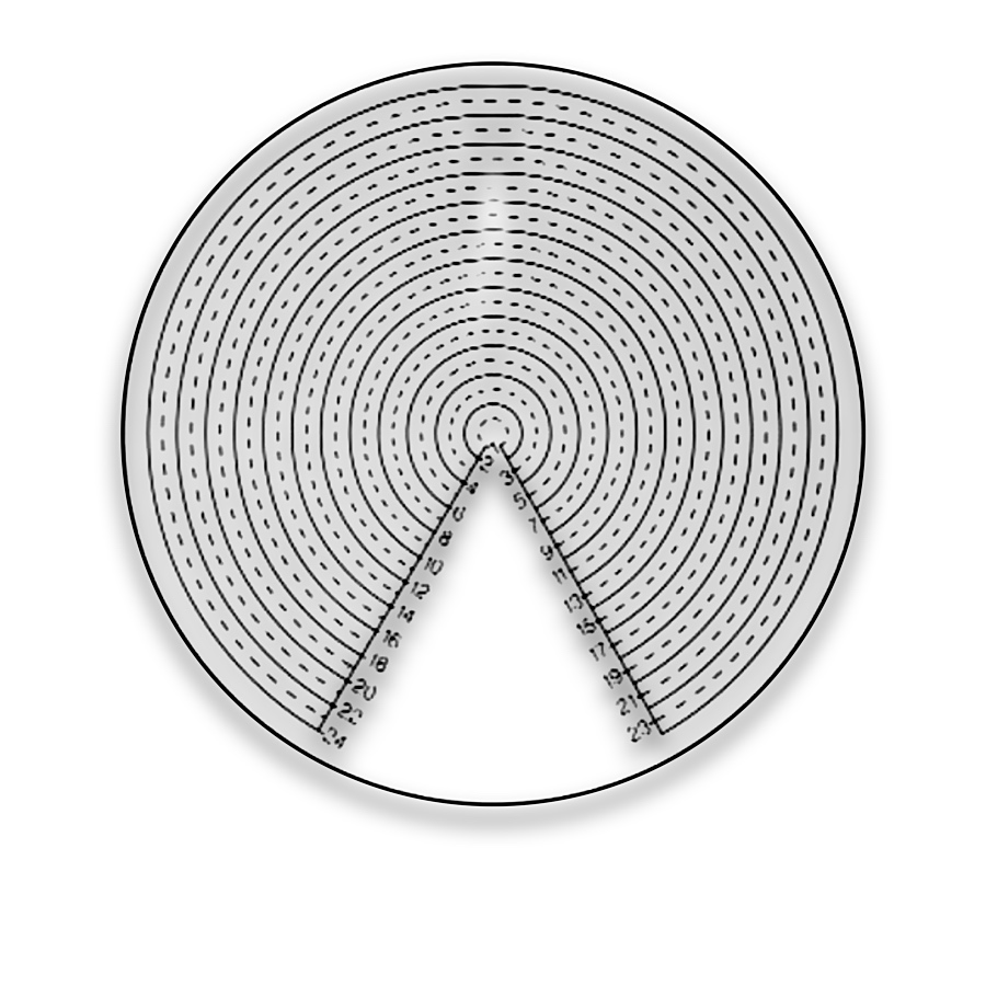
Comparison rings for circular features and particle sizes.
Angular divisions for orientation comparison.
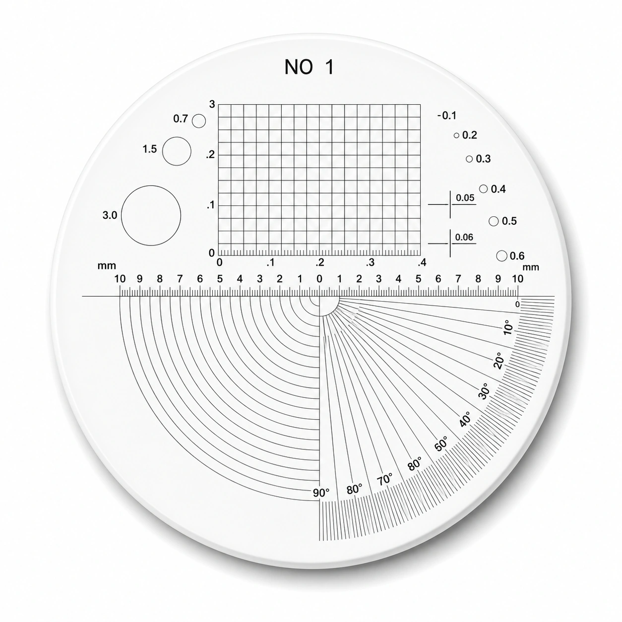
Multi-Scale Inspection Reticles for microscope measurement and optical inspection. Compare pattern layouts and choose the configuration for your instrument.
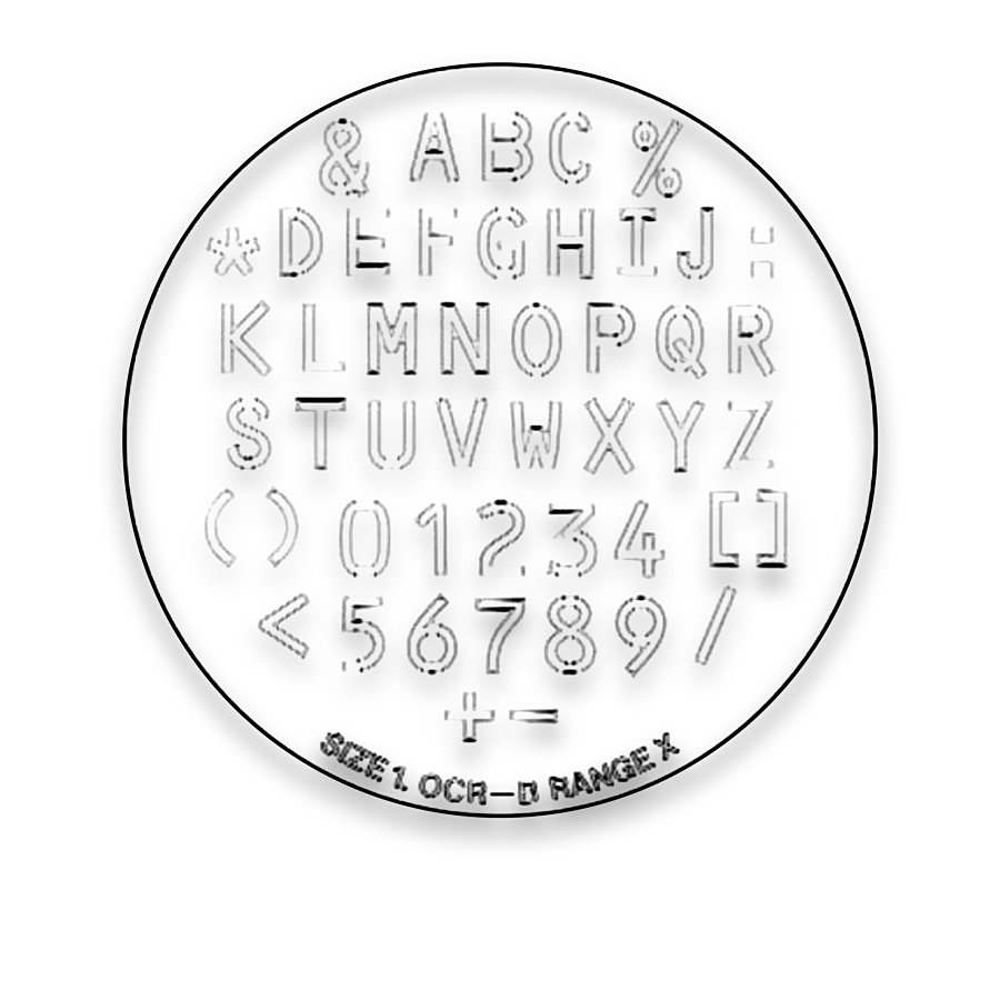
Character Comparison Reticles for microscope measurement and optical inspection. Compare pattern layouts and choose the configuration for your instrument.
Indexed polymer film for recording and relocating specimen regions.
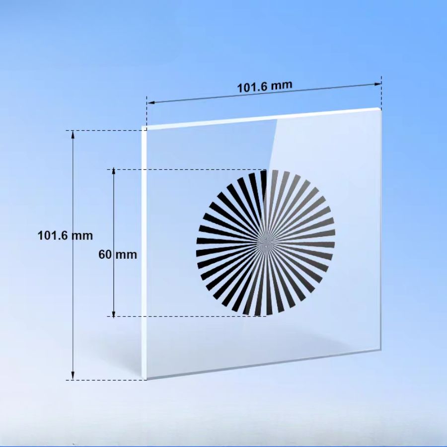
Radial detail for microscope focus and directional sharpness checks.
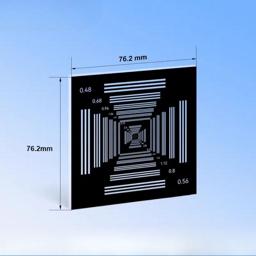
Three-bar groups for microscope focus and horizontal/vertical resolution checks.
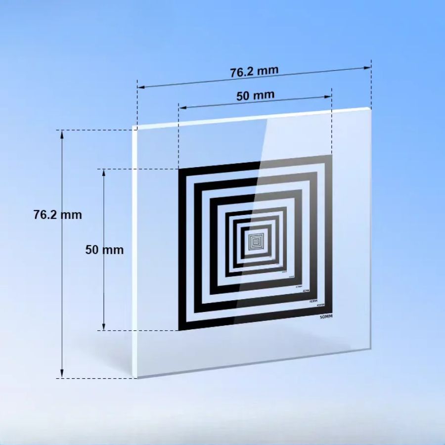
Known frame spans establish local image scale at the microscope specimen plane.
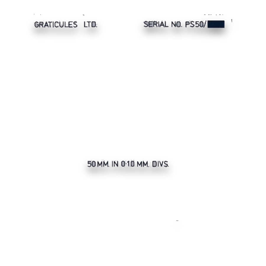
A known specimen-plane scale for eyepiece and camera calibration.
A stage micrometer provides a physical length reference at the specimen plane. An eyepiece reticle overlays graduations, grids or comparison shapes in the viewing system. Calibrating the reticle against a stage reference relates each division to a specimen dimension at the selected magnification.
Choose the pattern for the measurement or counting protocol, then confirm fit diameter, thickness and focal-plane location. Changing the objective or intermediate optics changes the calibration. Counting rules and sampling area belong to the method, while the reticle supplies the geometric reference.
Length measurement uses calibrated graduations. Counting uses an agreed observation area and boundary rule. Size comparison uses a defined reference series. Selecting these tasks separately helps avoid using a visually similar reticle with the wrong interpretation.
Stage micrometers establish a length reference at the specimen plane. Eyepiece scales and image-analysis systems use that reference to relate divisions or pixels to real dimensions. Recheck the relationship whenever the objective or optical configuration changes.
Counting grids divide a field into repeatable observation areas. Blood-cell and other concentration measurements also require a chamber with a defined depth, so the observed count can be related to sample volume. Laboratory cell suspensions, reproductive microscopy and food or water samples use different chambers and counting methods.
Comparison circles and graduated patterns help assess particle size, while fibre-counting graticules define a method-specific field. These references support dust examination, cleanliness inspection and pharmaceutical particle microscopy when matched to the required procedure and calibrated magnification.
Grain-comparison patterns, point arrays and test lines support grain-size examination, inclusion assessment and area-fraction sampling. Choose the reference geometry for the analysis method so repeated observations follow the same field, boundary and sampling rules.
An eyepiece reticle provides the visible reference; a stage micrometer establishes its specimen equivalent. Record the objective and intermediate magnification with the conversion. For two-axis measurements check both directions, and retain stage coordinates separately from eyepiece grid addresses.
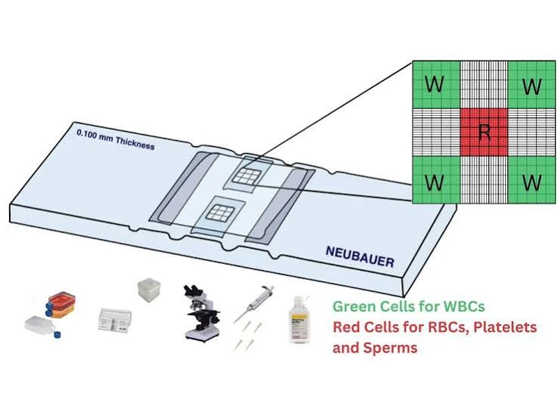
A Neubauer-style counting chamber illustrates how a defined observation grid supports cell counting. The chamber, cover and grid work together; the counting method and verified chamber geometry determine how a count is converted to concentration.
Microscopic measurement converts eyepiece divisions or image observations into lengths, size classes or counts.
Stage micrometers establish scale, reticles supply viewing references, and counting chambers additionally need matched depth and covers.
Length calibration, particle comparison, fibre counting and stereology need different patterns and methods. Select for the quantity being reported.