What the component does
The grid defines the area to be observed. An eyepiece counting reticle can help sampling, but does not by itself create a volumetric counting chamber.

Define a known observation volume so that a cell count can be related to the concentration of a prepared suspension.
Use the squares and boundary-counting convention specified by the method. The highlighted square represents an area, not a volume.
Counted volume = counted area × chamber depth. The chamber supports and seated cover establish the depth; the drawing is not to scale.
The grid defines the area to be observed. An eyepiece counting reticle can help sampling, but does not by itself create a volumetric counting chamber.
Use the chamber with its matching cover and filling method. Divide the count by the observed volume and apply the sample dilution factor where required.
An unsuitable cover, uneven filling, bubbles or the wrong depth assumption changes the effective volume and can bias the concentration result.
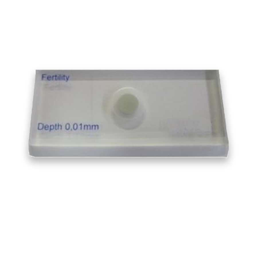
Explore the roles of the patterned base, chamber and cover.
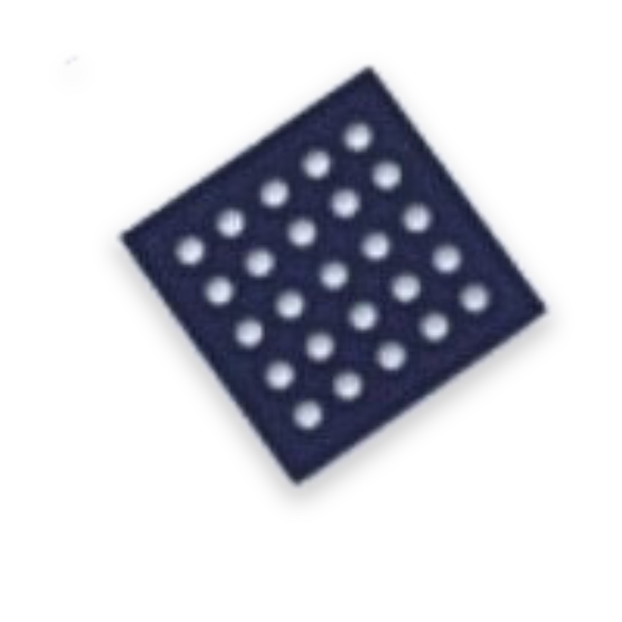
A matching cover seats on the chamber supports to establish the specified liquid depth.
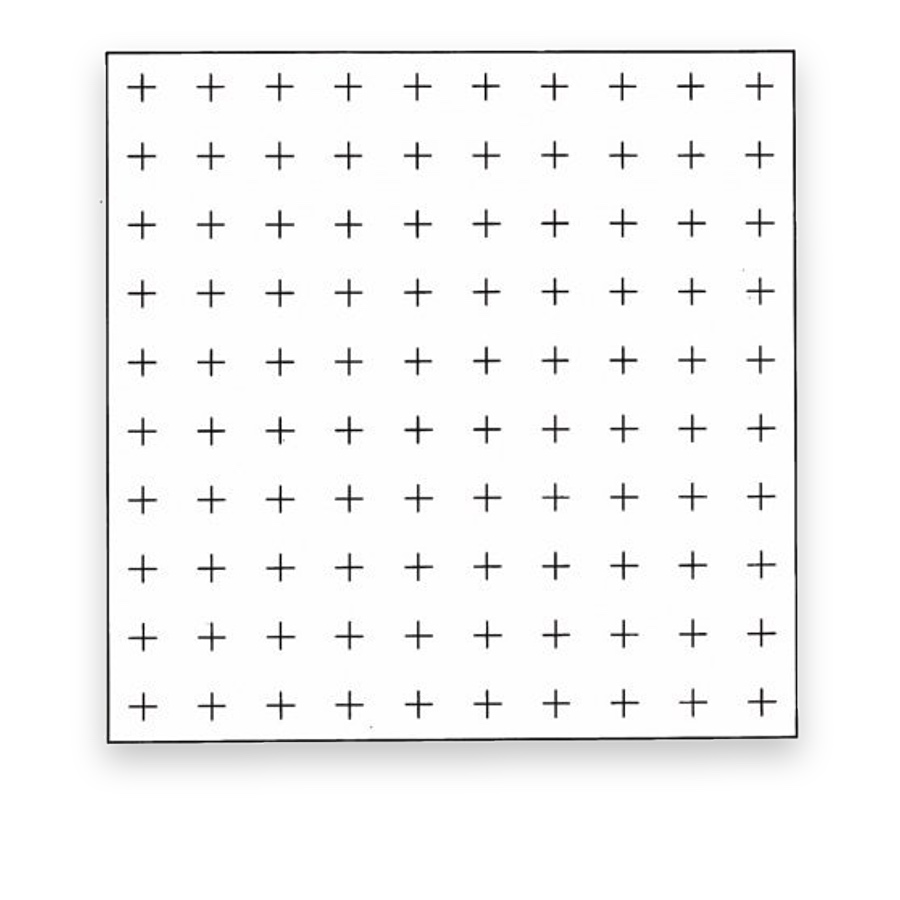
Provides a visual sampling grid; an eyepiece grid does not itself set chamber volume.
Length reference for checking the microscope image scale.
Microscopic concentration measurements relate the number of observed cells to a defined sample volume. In a haemocytometer, the counting area and chamber depth establish that volume. A patterned base, chamber structure and cover work together; an eyepiece grid alone supplies only a visual sampling area.
Choose the chamber and grid required by the specimen and counting method. Improved Neubauer, Thoma and other layouts differ in their subdivisions and conventions. Select the grid and chamber geometry together, then apply the boundary and counting rules belonging to that method.
Relevant settings include laboratory cell suspensions, specified manual blood-cell methods and method-specific reproductive microscopy. These operations take place after sample collection and preparation. The patterned glass helps observation and counting; it is not the blood-collection device.
The effective volume depends on correct cover seating and the defined separation from the chamber floor. Uneven filling, bubbles or an unsuitable cover can change the observation conditions even if the grid lines are correct. Depth and grid-area verification are different checks.
Some bright-line chamber designs use a coated background with clear ruling so the grid can be seen with suitable illumination. Other products use different ruling constructions. Coating polarity describes visibility; it does not establish chamber depth, biocompatibility or a complete diagnostic specification.
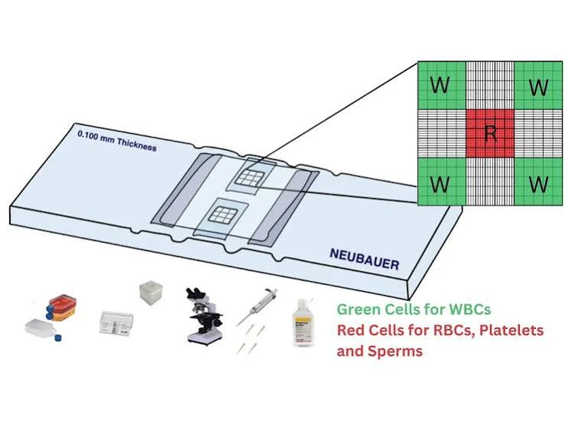
A Neubauer-style counting chamber illustrates how a defined observation grid supports cell counting. The chamber, cover and grid work together; the counting method and verified chamber geometry determine how a count is converted to concentration.
Cell-counting chambers use a grid, chamber depth and properly seated cover to define observation geometry.
Apply the selected preparation and counting method, then use known volume and dilution to calculate concentration.
Volume also depends on area, depth and filling. Bubbles, unmatched covers and dilution or counting records can change the calculation.