
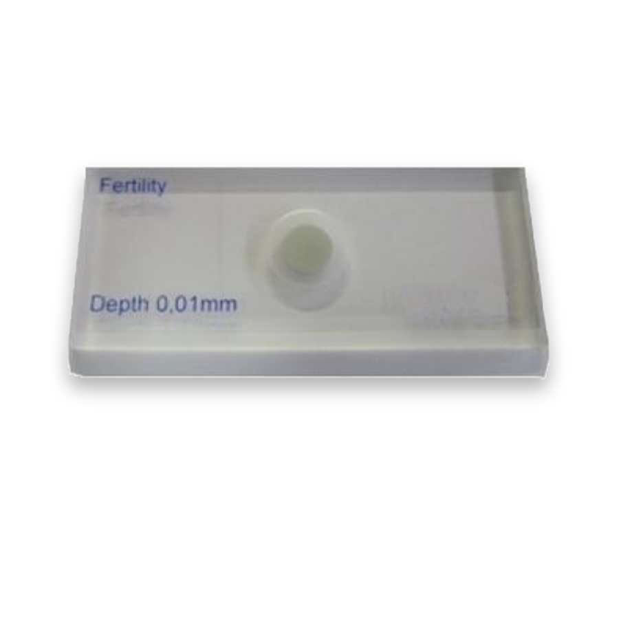
Microscopy Counting Chamber Components

A counting chamber component supplies part of the geometry used to observe a liquid sample. The patterned floor, chamber supports and matching cover establish the counted area and depth; the supplied fertility-analysis examples belong to that chamber family.

Microscopy Counting Chamber Components

Configuration and materials

Specify glass grade, dimensions, thickness, surface quality and the mating chamber geometry. Any complete assembly needs defined seating, chamber depth, cleaning compatibility and an agreed verification procedure.

  • Active field, scale or feature geometry and optical-plane location
  • Material, outside dimensions, thickness and mounting datums
  • Working instrument, illumination and viewing side
  • Critical tolerances, inspection intervals and reporting needs

Reading and sampling methods

Grid, chamber and cover form one volume

The grid fixes the area being counted; the chamber support and correctly seated cover establish the depth. These dimensions must belong to the same chamber design before a count can be converted to concentration.

Calculate counting volume and concentration

Match the chamber to the method

Blood-cell, fertility-analysis and organism-counting chambers use different depths, fields and preparation methods. The pictured fertility-analysis reference does not define the geometry of a Neubauer blood-cell chamber.

Choose the pattern for the counting method

Choose by function

What it is and what it does

A counting chamber component supplies part of the geometry used to observe a liquid sample. The patterned floor, chamber supports and matching cover establish the counted area and depth; the supplied fertility-analysis examples belong to that chamber family.

When to choose it

Choose a counting chamber when the result depends on the number of objects in a known liquid volume. The chamber family must suit the specimen and loading method.

How it differs from related products

A counting chamber combines a defined area with a defined depth. Its cover is part of that geometry. A generic grid, ordinary cover glass or differently sized chamber does not preserve the same counted volume.

What can be customised

Choose chamber family, grid layout and matching cover geometry for the specimen method. Howard cells, Sedgewick–Rafter chambers and haemocytometers serve different workflows.

What you receive

The configuration must identify the chamber body, counting pattern and matching cover as separate supplied items. The pattern illustrations identify the chamber family; a diagnostic kit, validated assay and instrument are outside that description.

Where this product is used

Pattern and format options

Microscopy Counting Chamber Components — Pattern 066

Pattern 066

Related pattern choices

Selection notes

Microscopy counting-chamber components combine an observation grid, chamber and matching cover to define a specimen layer.

Specify grid geometry and the chamber-cover interface together. The counting method uses both the observed area and chamber depth.

Information for your inquiry

Grid pattern, chamber depth, seating geometry, cover format, microscope setup and inspection requirements.

Why must the cover be matched to the chamber?

The cover and support faces form the liquid layer. Different seating or geometry changes the observation volume used in the calculation.

Search