What the component does
Patterned slides and fiducials help locate fields or revisit sample positions. A stage micrometer provides a separate length reference for image scale.

Provide spatial references for laboratory imaging so that fields, sample positions and image scale can be checked consistently.
Reference and sample positions are illustrated together; actual designs may use separate calibration and sample slides.
These components support instrument referencing; they do not by themselves establish clinical diagnostic performance.
Patterned slides and fiducials help locate fields or revisit sample positions. A stage micrometer provides a separate length reference for image scale.
Optics, sample support and software coordinate conventions need to agree. If counting concentration, use a chamber with a defined volume rather than only an image grid.
A reference at a different sample height can change apparent scale or focus. Pattern chemistry and cleaning compatibility must suit the actual instrument workflow.
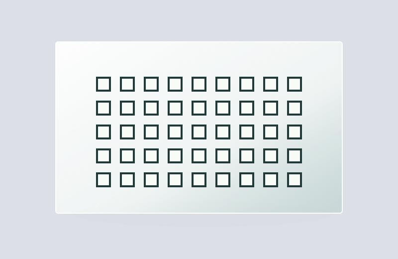
Repeated sites and viewing windows for instrument scan registration.
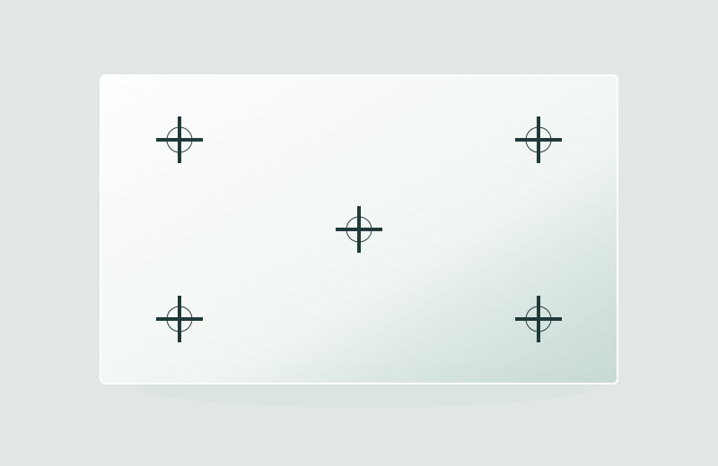
Reference marks to align repeated images or slide positions.
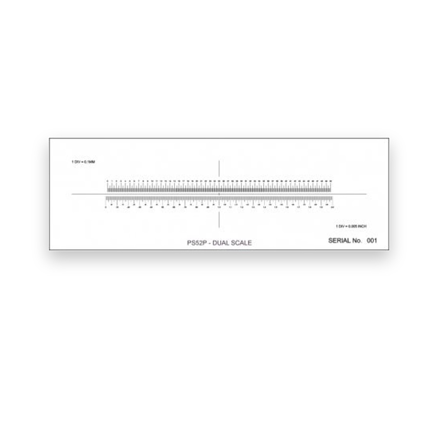
Specimen-plane length references for image calibration.
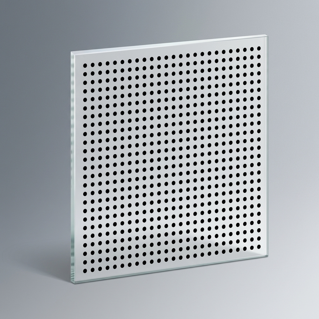
Camera-coordinate references across a defined field.
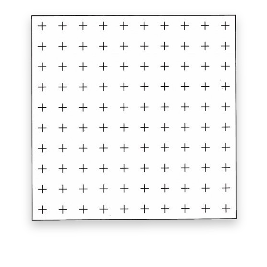
Geometric field subdivisions for a defined counting workflow.
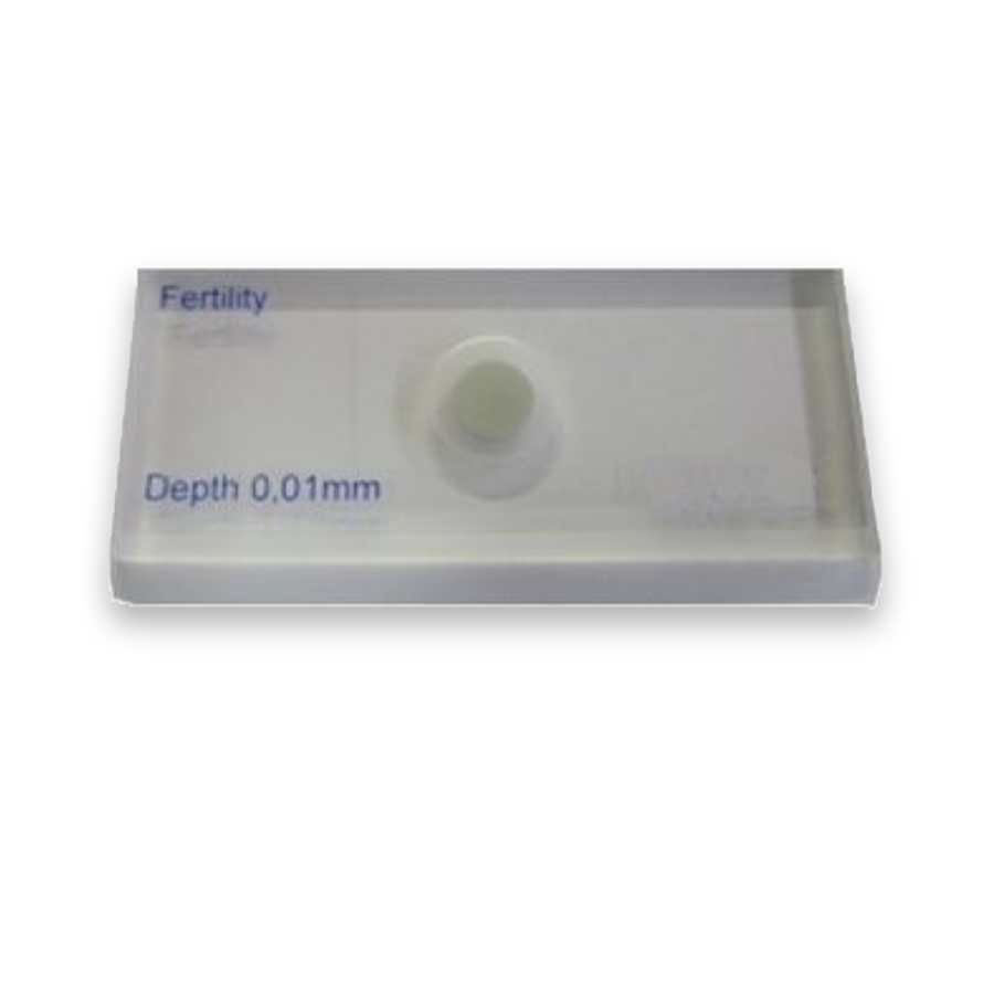
Patterned-base and chamber considerations for method-specific cell observation.
Laboratory instruments may need stable feature coordinates, repeatable scan alignment and a known image scale. A patterned slide can locate imaging sites, while calibration targets check the camera geometry or pixel-to-length relationship. Eyepiece patterns can support a defined visual counting method.
Select an optical reference that matches the instrument workflow. Patterned windows do not by themselves create fluid wells or biological assay chemistry. Surface contact, cleaning, illumination and any instrument-specific validation should be defined independently of the geometric pattern.
An instrument may use a calibration target to check camera geometry, a scale to verify image dimensions and a counting chamber to observe a specimen. The first two check the optical measurement chain; the last also depends on specimen preparation and chamber configuration.
Reference glass in laboratory and diagnostic imaging supports scale checks, region location and method-specific microscopic observations.
Define the optical reference separately from sample handling, assay chemistry and complete-instrument validation.
A film pattern may provide only an optical window or location mark. Liquid chambers and wells require corresponding physical structures.