Eyepiece Micrometer Reticles

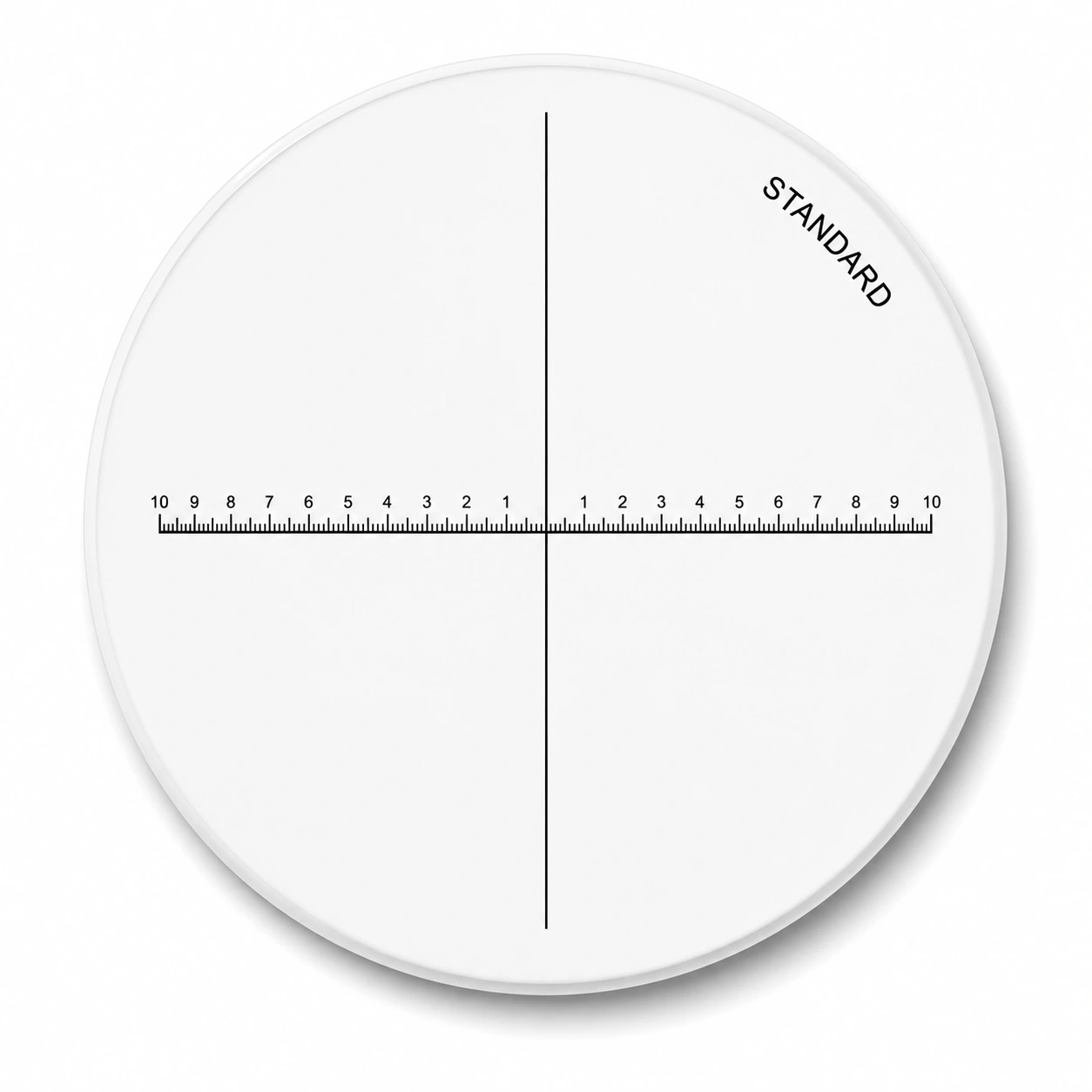
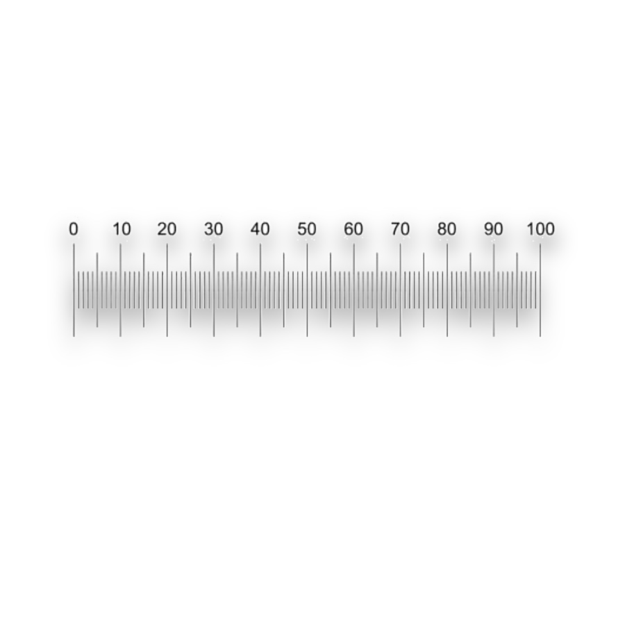
An eyepiece micrometer reticle places a graduated reference in the microscope image plane. It lets an operator measure particle lengths, fibre widths and small component features against a scale visible with the specimen.

Eyepiece Micrometer Reticles

Configuration and materials

Specify optical glass or fused silica, overall dimensions, thickness, pattern-side orientation and the clear aperture. Define transmission, reflectance or optical density at the working wavelength when optical contrast is critical.

  • Reticle seat diameter and thickness clearance
  • Scale length, graduation, line width and numbered intervals
  • Pattern plane, reading side and single-axis or XY layout

From the pattern to a measurement

A scale at the eyepiece image plane

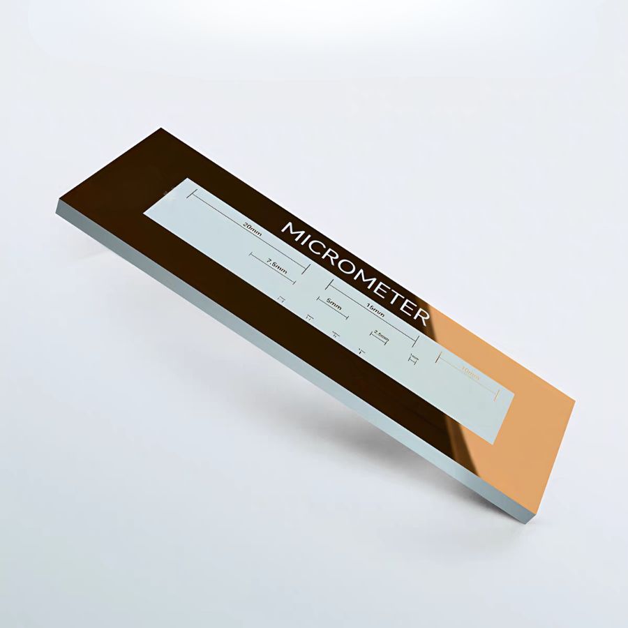
The observer compares a feature with fixed divisions without moving a physical ruler onto the specimen. A stage micrometer establishes the specimen length represented by each division for the objective in use.

Calibrate eyepiece divisions against a stage scale
Measurement range and readable detail

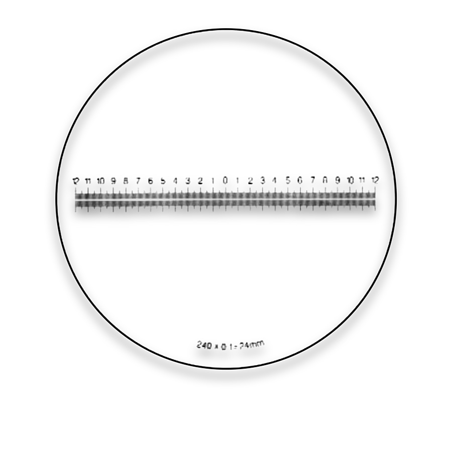
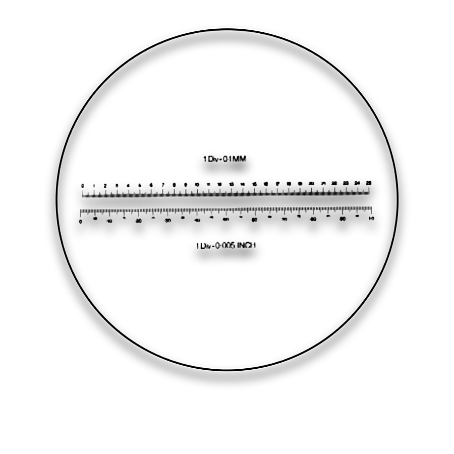
Graduated length controls coverage and pitch controls the available reference intervals. Finer marks can be harder to resolve at low contrast; line width and numbering should leave the specimen visible.

Specify contrast and viewing side

Choose by function

What it is and what it does

An eyepiece micrometer reticle places a graduated reference in the microscope image plane. It lets an operator measure particle lengths, fibre widths and small component features against a scale visible with the specimen.

When to choose it

Choose a single scale for measurements along one direction. Choose crossed scales for two perpendicular directions or for measurements that must share a common origin. Rotate the reference or specimen so the measured feature follows the scale axis.

How it differs from related products

An eyepiece division has no fixed specimen value until calibrated. A stage micrometer supplies a known length at the specimen plane; a crosshair without graduations supplies an alignment reference, not a length scale.

What can be customised

Scale length, graduation interval, numbering, line width and a single or crossed layout define the visible reference. Disc diameter, thickness, seating face and pattern position define its fit in the eyepiece.

What you receive

A patterned reticle disc for the eyepiece or measuring magnifier. Its physical scale geometry is separate from the specimen calibration established on the assembled microscope.

Where this product is used

Pattern and format options

Eyepiece Micrometer Reticles — NE1

NE1

  • The supplied designation identifies a nominal 10 mm scale with 0.1 mm divisions at the reticle plane. These dimensions do not equal specimen-plane distances until the microscope is calibrated.
Eyepiece Micrometer Reticles — M6T10

M6T10

Eyepiece Micrometer Reticles — M6T20

M6T20

Eyepiece Micrometer Reticles — M6T1

M6T1

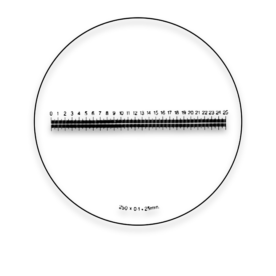
Eyepiece Micrometer Reticles — Micrometer Scale Reticle

Micrometer Scale Reticle

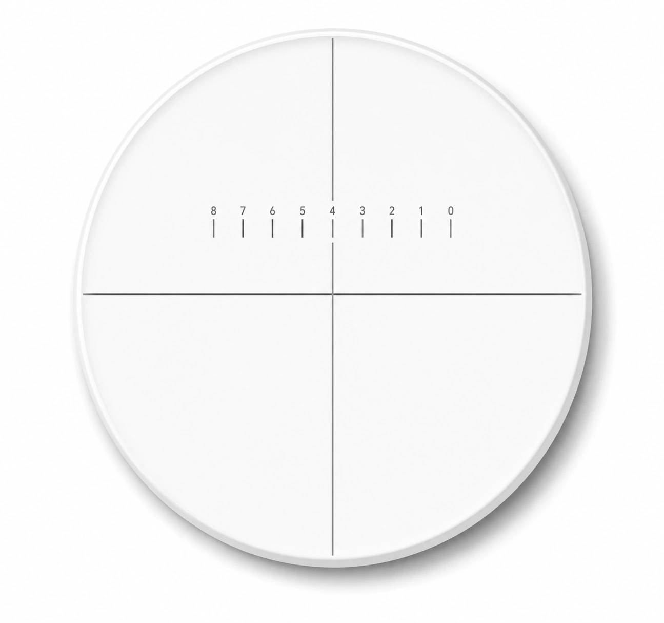
Eyepiece Micrometer Reticles — Crosshair with a numbered scale

Crosshair with a numbered scale

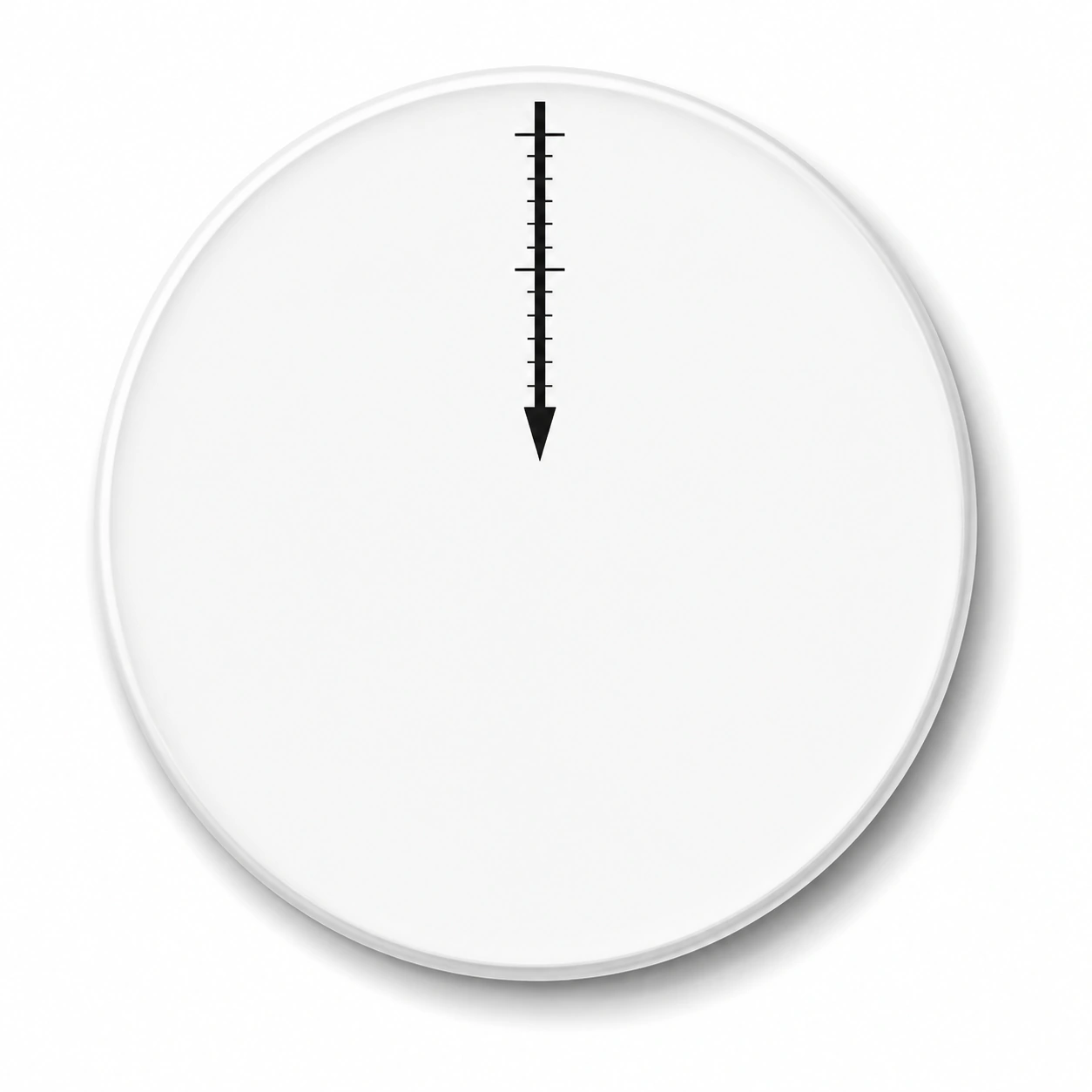
Eyepiece Micrometer Reticles — Graduated pointer scale

Graduated pointer scale

Eyepiece Micrometer Reticles — Horizontal scale with a reference axis

Horizontal scale with a reference axis

Common questions

Does a 0.1 mm reticle division mean 0.1 mm on the specimen?

No. It is a physical distance in the reticle plane. Its specimen equivalent depends on the optical magnification and is established with a stage reference.

Will one calibration cover every objective?

Keep a separate conversion for each objective and intermediate optical configuration. Nominal objective labels alone do not capture every magnification change.

Is the eyepiece diameter enough to choose a reticle?

Measure the reticle seat, thickness clearance and usable pattern field. The outside diameter of the eyepiece housing is not the reticle diameter.

Related pattern choices

Technical reading

Selection notes

Micrometer reticles introduce graduations into the viewing field for comparing lengths, spacing and displacement.

Define division spacing, scale length, numbering and reading direction. Calibrate the specimen value of each division in the actual optical configuration.

Information for your inquiry

Division pitch, total scale length, numbering, eyepiece fit, magnification and film side.

Does each division always represent the same specimen length?

The specimen scale changes with objective, zoom and optical configuration. Establish it with a stage micrometer and record the setup.

Search